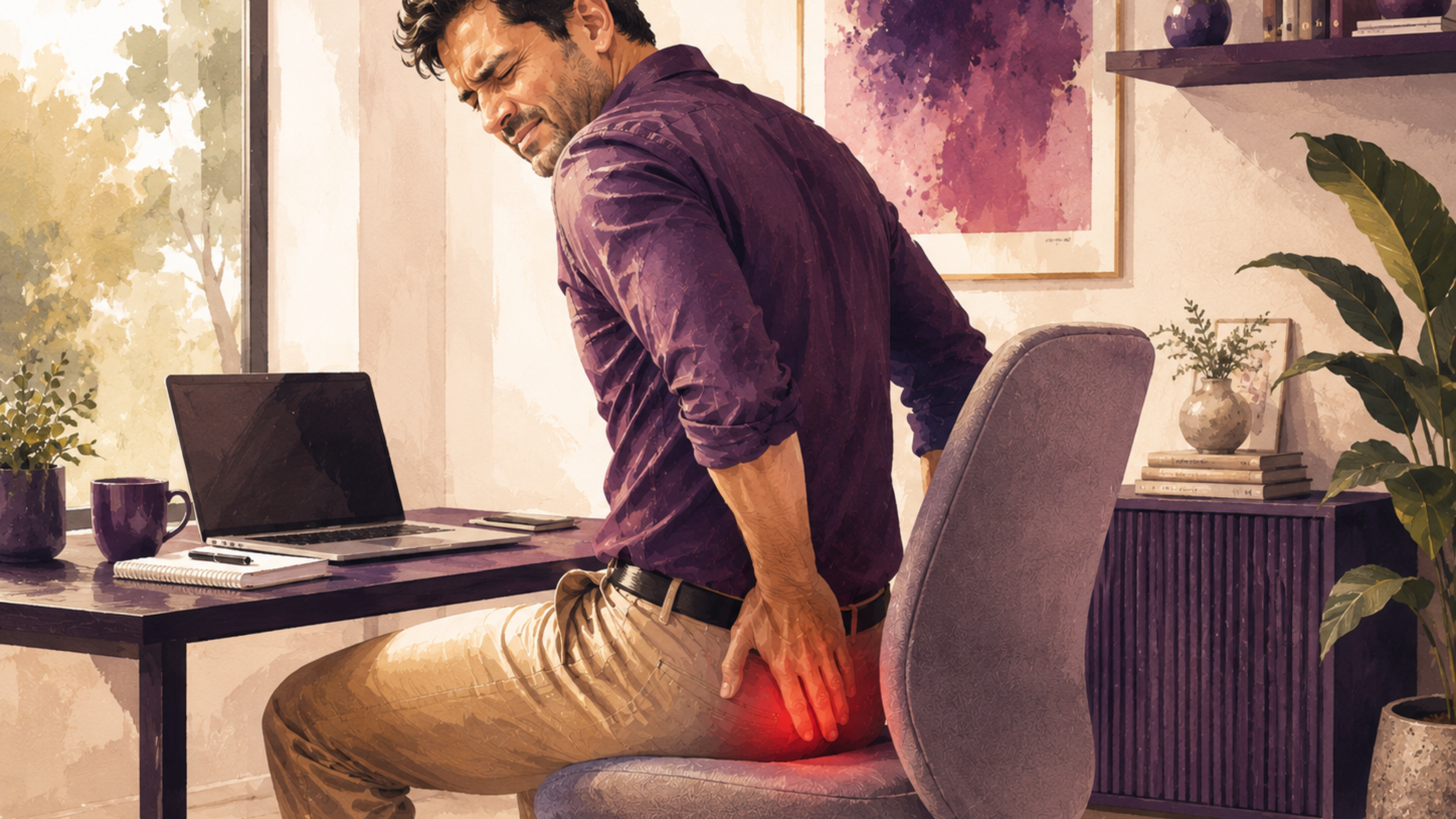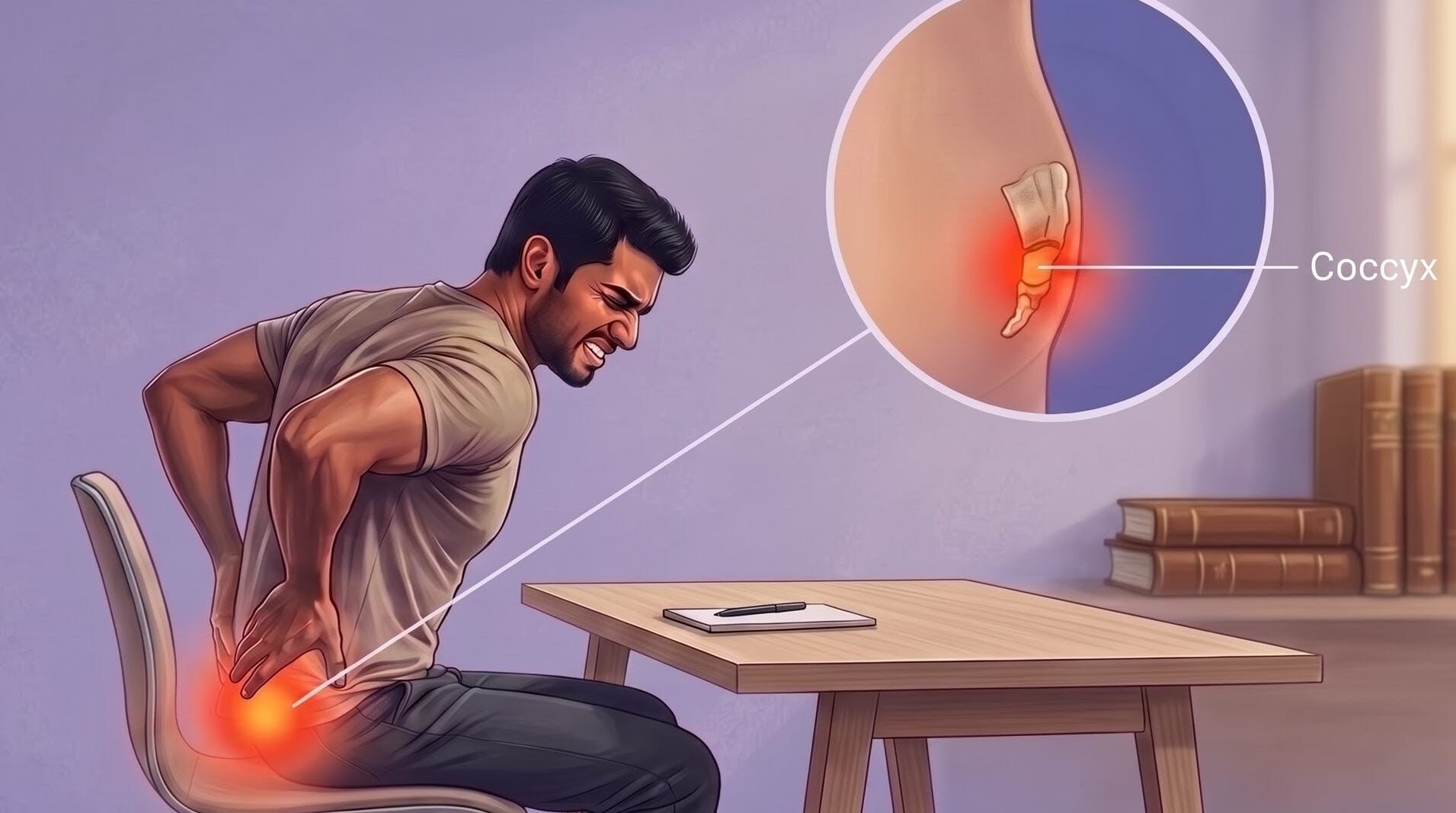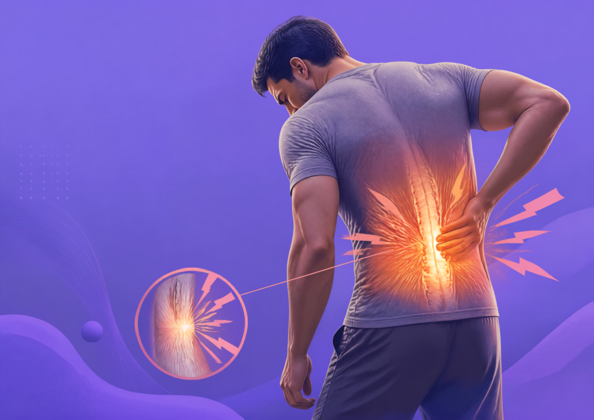




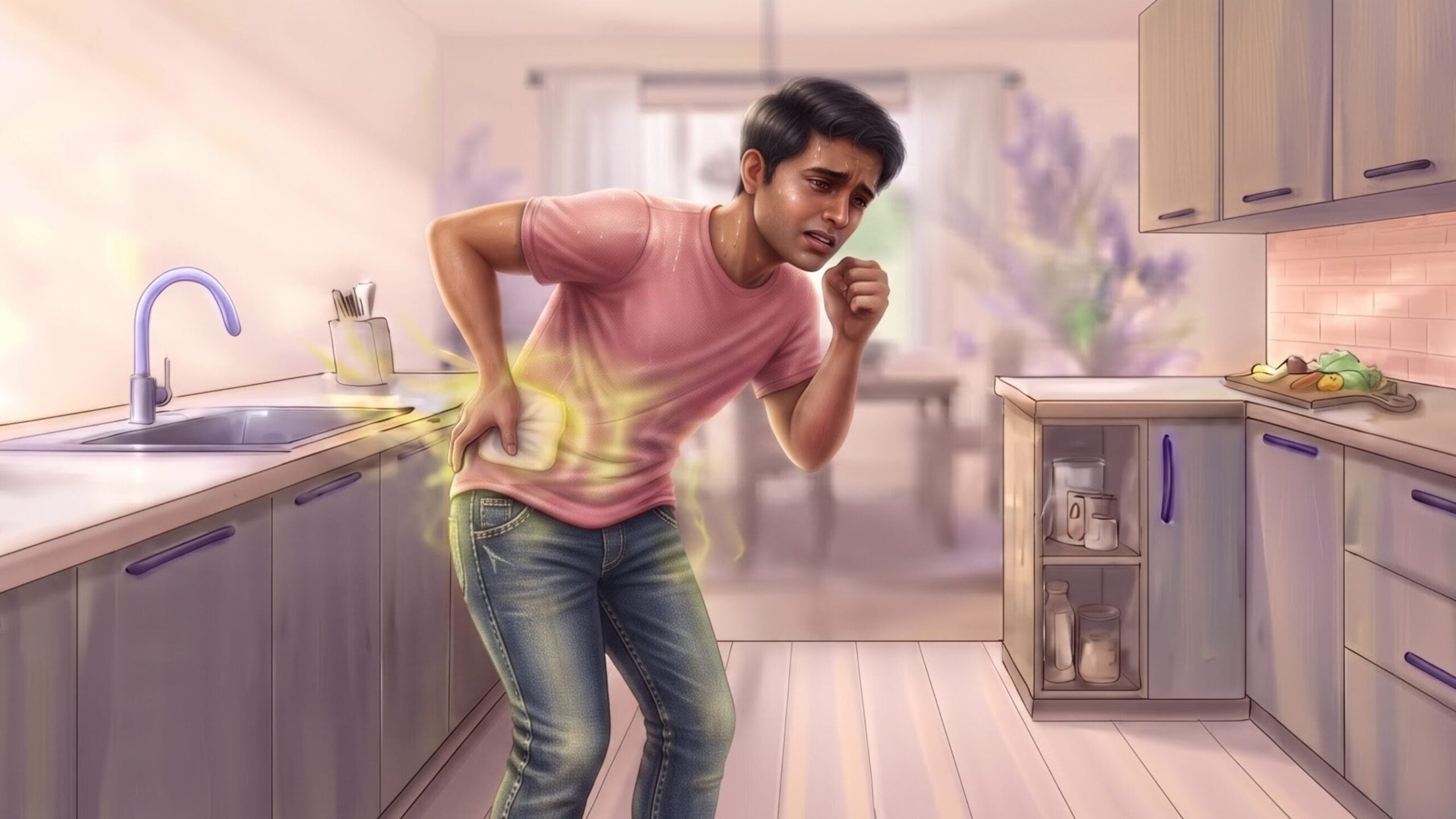
Experiencing sudden back pain after a minor movement like coughing, reaching, or lifting a light package can be confusing and alarming [1]. While many people attribute unexpected back pain to a pulled muscle or a bad disc, sharp pain triggered by low-load everyday activities can sometimes point to a structural change in the spine known as a vertebral compression fracture (VCF) [1].
This guide explains what happens during a vertebral compression fracture, how underlying bone mineral density plays a role, key signs to look for, and how healthcare providers evaluate and manage the condition safely [1, 2].
What Is a Vertebral Compression Fracture?
A vertebral compression fracture happens when a bone in your spine partially collapses under weight or pressure [1]. The human spine is made up of individual bones called vertebrae stacked on top of one another. Each vertebra features a dense, hard outer shell surrounding a softer, honeycomb-like interior called trabecular bone (or cancellous bone) [1].
When this sponge-like inner bone matrix weakens, it can no longer support everyday weight-bearing forces [1, 2]. Under axial pressure, which is downward force along the length of your spine, the interior structure develops tiny micro-fractures, causing the front portion of the spinal bone to compress and collapse downward [1].
This collapse most commonly affects the thoracic spine (the middle back) and the thoracolumbar junction (the transitional area where your middle back meets your lower back) regions naturally subjected to significant physical stress during daily movement [1].
How Osteoporosis Weakens Vertebral Bone
Osteoporosis is a systemic bone disease that causes bones to lose density and structural strength [2]. As bone density declines a process accelerated in post-menopausal individuals due to hormonal shifts, as well as in older adults, the internal scaffolding of the spinal bones becomes porous and fragile [2].
When bone remodeling (the body’s natural balance of breaking down old bone tissue and rebuilding new bone) falls behind, the inner support beams inside your spine become thin and brittle [2]. This bone loss (or its milder precursor, osteopenia, which means lower-than-normal bone density) significantly reduces the mechanical force a vertebra can tolerate before failing [2].
Why Everyday Movements Can Trigger Structural Collapse
In a healthy spine, high-energy trauma like a car crash or a fall from a significant height is usually required to break a spinal bone [1]. However, when osteoporosis severely weakens bone mineral density, the spine can develop fragility fractures breaks caused by low-impact, routine activities [2].
When the internal bone matrix is fragile, ordinary forces generated by daily movements can exceed what the bone can support [1, 2]. Common triggers include:
- Bending forward to pick up a light item from the floor [1].
- A sudden, severe fit of coughing or sneezing [2].
- Reaching up toward an overhead shelf [1].
- Turning or twisting abruptly in bed [1].
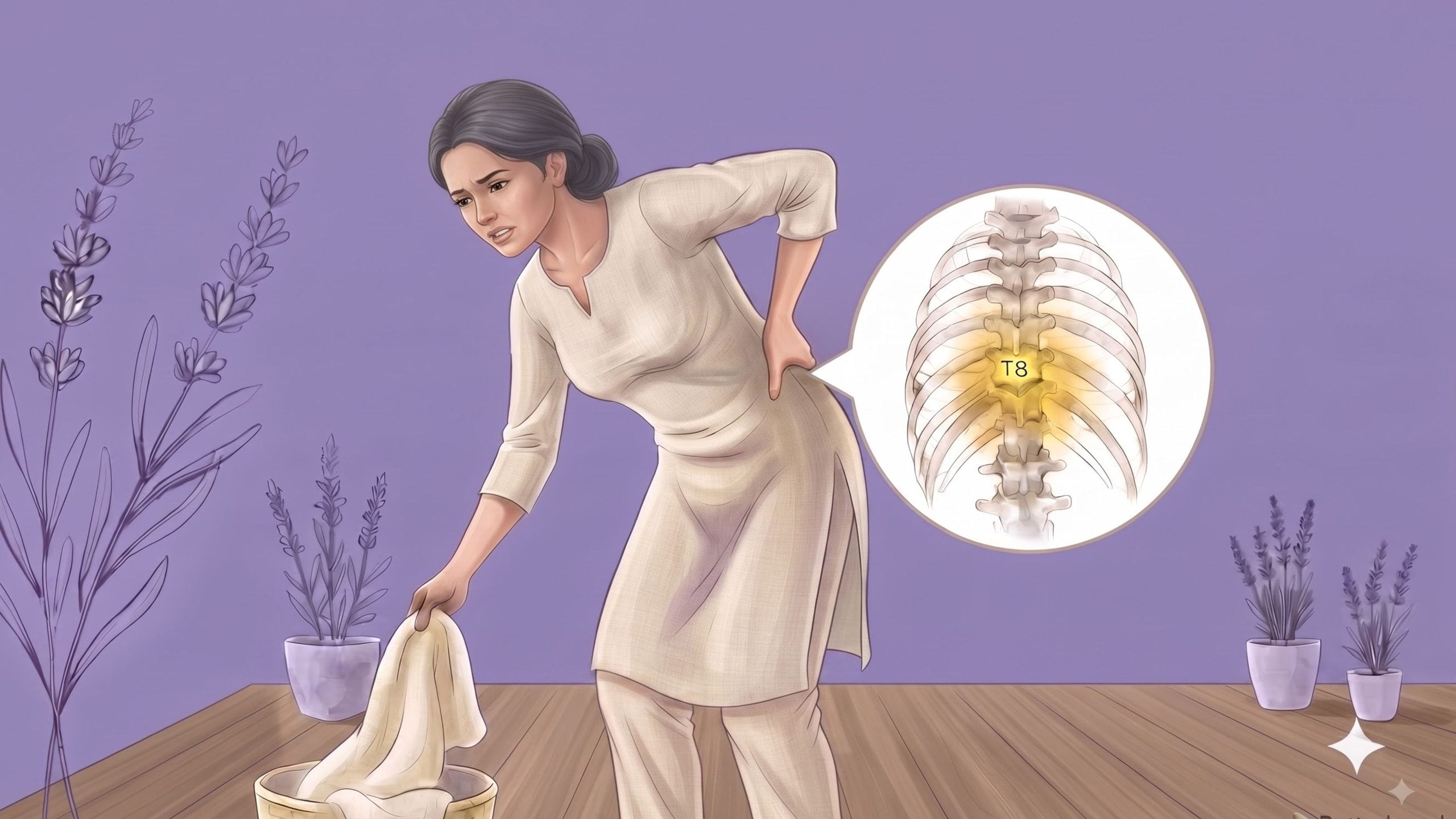
Signs and Symptoms of a Vertebral Compression Fracture
Vertebral compression fractures can present differently depending on whether the structural collapse happens suddenly or develops gradually over time [1].
Sudden Axial Back Pain vs. Chronic Aches
The most common sign of an acute (recent) compression fracture caused by bone loss is the sudden onset of sharp, localized axial back pain meaning pain centered directly along the spine rather than spreading down the legs [1].
- Acute Pain: Patients often report a sudden “snapping” or sharp aching sensation immediately following a low-impact movement [1].
- Postural Sensitivity: Pain typically worsens during weight-bearing activities, such as standing, walking, or sitting upright for extended periods [1].
- Positional Relief: Back pain from a compression fracture often improves significantly when lying flat on your back (the supine position), because lying down removes gravitational pressure from the compressed spinal bone [1].
- Localized Tenderness: Pressing directly over the affected spinous process (the bony point you can feel along the center of your back) often reproduces distinct tenderness [1].
In contrast, some compression fractures develop slowly without a single, dramatic painful event [1]. Individuals may experience a dull, persistent back ache that is easily mistaken for general muscle fatigue or age-related stiffness [1].
Height Loss, Slouching, and Progressive Kyphosis
A compression fracture usually causes the front of the spinal bone to collapse. The denser back wall often remains largely intact, creating a wedge shape [1].
When one or more spinal bones collapse into this wedge shape, posture and spinal alignment can change [1, 2]:
- Progressive Kyphosis: Forward wedging can tilt the spine forward. This may contribute to kyphosis, an exaggerated rounding of the upper back [1].
- Measurable Height Loss: As individual spinal bones lose vertical height, a person’s total standing height decreases over time [2].
- Postural Changes: The shoulders may round forward as the spine changes. The ribcage may also settle closer to the pelvis. This can cause abdominal discomfort or a crowded feeling in the torso [1].
How to Tell a Compression Fracture from Other Back Pain
Because back pain is extraordinarily common, distinguishing a vertebral fracture from soft-tissue injuries or disc conditions is critical for getting the right medical care [1].
Comparing Fractures, Muscle Strains, Disc Disease, and Stenosis
| Condition | What usually makes it different |
| Vertebral Compression Fracture (VCF) [1] | Sharp pain centered directly along the spine that worsens when standing and eases when lying flat; may cause height loss or slouching. |
| Lumbar Muscle Strain [1] | Dull aching or tightness in back muscles triggered by bending or moving, without height loss or bone shape changes. |
| Degenerative Disc Disease (DDD) [1] | Persistent stiffness and deep aching in the spine that typically feels worse during prolonged sitting. |
| Lumbar Spinal Stenosis [1] | Leg pain, heaviness, or cramping when walking or standing upright that improves when leaning forward. |
How Vertebral Compression Fractures Are Diagnosed
A thorough clinical evaluation involves confirming whether a fracture exists, assessing how recent it is, and evaluating your overall bone health [1, 3].
Spinal Imaging: Confirming Acute Fractures with X-rays and MRI
When a healthcare provider suspects a vertebral fracture, structural spinal imaging is the first step [1, 3]:
- Standard X-rays: Plain radiographs (front and side views) show the shape and height of your spinal bones. X-rays can easily confirm front-wall wedging, height loss, and changes in overall spinal curvature [1].
- Magnetic Resonance Imaging (MRI): X-rays show bone structure but cannot easily distinguish a brand-new, actively healing fracture from an older, stable one. An MRI detects bone marrow edema swelling and fluid buildup inside the bone that marks a recent or acute fracture [3].
- Computed Tomography (CT) Scans: Used when an MRI cannot be performed or when a doctor needs a detailed, cross-sectional view of the hard bony architecture to evaluate structural stability [1].
DEXA Scans: Evaluating Underlying Bone Mineral Density
While X-rays and MRIs evaluate the structural break, a DEXA scan (Dual-Energy X-ray Absorptiometry) a low-dose X-ray scan that measures bone mineral density evaluates how dense and strong your bones are overall [2].
A DEXA scan compares your bone density to that of a healthy young adult, yielding a T-score [2]:
- Normal: T-score of -1.0 or higher [2].
- Osteopenia (Low Bone Mass): T-score between -1.0 and -2.5 [2].
- Osteoporosis: T-score of -2.5 or lower [2].
Diagnosing underlying bone loss is critical because experiencing a single fragility fracture significantly increases the likelihood of suffering subsequent fractures in the future [2, 5].
Nonsurgical Recovery and Safe Movement
The majority of uncomplicated vertebral compression fractures achieve substantial pain relief and functional stability through conservative, nonsurgical management under medical supervision [1, 4].
Daily Movement Precautions: Protecting a Healing Spine
During early recovery, protect the spine from excessive loading and forward bending. This can help support comfort and healing [1].
- Avoid Heavy Flexion Under Load: Avoid movements that involve heavy forward bending during early recovery. Examples include weighted toe-touches, heavy lifting, and aggressive sit-ups. Follow your clinician’s guidance before resuming these activities [1].
- Maintain Neutral Alignment: Move your body as a single unit when changing positions. Use the “log-rolling” technique when turning in bed or getting up. Keep your spine in a tall, neutral position [1].
- Avoid Heavy Lifting: Avoid carrying heavy items or pulling heavy loads overhead. Also avoid twisting while holding objects. These movements can place extra strain on vulnerable vertebrae [1].
- Gradual, Safe Activity: Short periods of rest may help relieve severe pain at first. However, prolonged bed rest can contribute to bone loss and muscle weakness. Controlled, light walking on flat surfaces is usually encouraged as tolerated to support systemic bone health [1, 2].
The Role and Limits of Medical Bracing
In certain clinical scenarios, an orthopedist or spine specialist may prescribe a specialized back brace [1, 4].
- Rigid/Hyperextension Orthoses (e.g., TLSO): A Thoracolumbar Sacral Orthosis is a rigid brace designed to support the middle and lower spine while holding you in a straight, upright position. This takes pressure off the front column of the spinal bone, helping reduce pain during movement [1, 4].
- Limits of Elastic Belts: While elastic support belts can offer mild compression and postural awareness, they do not provide rigid mechanical stabilization or heal a fracture. A spine specialist will determine whether a rigid orthosis or a lighter support belt is appropriate based on your fracture’s stability [1, 4].
Medical Management and Nonsurgical Pain Relief
Nonsurgical care focuses on managing symptoms so you can stay safely mobile [1, 2]:
- Medications: Physicians may recommend short-term over-the-counter pain relievers, prescription analgesics, or short courses of anti-inflammatory medications, tailored to your overall health and kidney function [1].
- Targeted Bone Therapies: In acute settings, specific bone-preserving medications or hormonal sprays (such as calcitonin) are sometimes prescribed by physicians to help ease acute bone pain [2].
- Physical Therapy: Once initial acute pain settles, a physical therapist can guide gentle posture training, balance exercises, and safe core stabilization techniques focused on back extension [1].
How to Prevent Future Vertebral Fractures
A primary goal after diagnosing a compression fracture is stopping the “fracture cascade” a cycle where one collapsed vertebra shifts mechanical load to adjacent spinal bones, increasing their risk of failure [2, 5].
Preserving Bone Health and Mineral Density
- Osteoporosis Pharmacotherapy: Medical providers often recommend bone-preserving medications such as bisphosphonates, RANKL inhibitors (targeted therapies that prevent bone breakdown), or bone-building anabolic agents to improve bone density and reduce future fracture risk [2].
- Nutritional Support: Ensuring adequate intake of Calcium and Vitamin D supports healthy bone remodeling. Nutritional approaches should always be discussed with a doctor to tailor dosing to your individual blood levels and health conditions [2].
- Weight-Bearing Exercise: Regular, low-impact, weight-bearing activities (such as walking) along with gentle resistance training stimulate osteoblasts (cells that help build new bone) to help preserve bone mass [2].
Fall Prevention and Home Safety
Because unexpected trips or slips can subject a fragile spine to sudden impact, fall-proofing your living environment is an essential preventive measure [2]:
- Clear Walkways: Remove loose throw rugs, electrical cords, and low coffee tables from primary walking paths [2].
- Improve Lighting: Ensure hallways, stairwells, and pathways between the bedroom and bathroom are well-lit, especially at night [2].
- Install Grab Bars: Place sturdy handrails along stairways and secure grab bars inside showers and beside toilets [2].
- Footwear Matters: Wear non-skid, supportive footwear indoors rather than walking in bare feet, smooth socks, or loose slippers [2].
When to Seek Urgent Medical Evaluation
While most vertebral compression fractures are managed conservatively, sudden back pain accompanied by specific warning signs requires prompt or emergency medical assessment [1].
Seek immediate medical attention if back pain is accompanied by any of the following symptoms [1]:
- Neurological Deficits: Symptoms like numbness, tingling, or progressive weakness in your legs, feet, or groin area [1].
- Bowel or Bladder Dysfunction: Sudden loss of bowel or bladder control, or new difficulty urinating [1].
- Saddle Anesthesia: Loss of sensation in the areas that would touch a saddle (your inner thighs, buttocks, and groin) [1].
- High-Energy Trauma: Back pain resulting from a major fall, car crash, or direct heavy impact [1].
- Systemic Signs: Unexplained fever, chills, night sweats, or unexpected weight loss accompanying new spinal pain [1].
Frequently Asked Questions (FAQ)
What does a vertebral compression fracture feel like?
An acute vertebral compression fracture usually causes sudden, sharp back pain centered directly along the spine [1]. The pain typically intensifies when standing, walking, or bending, but improves significantly when lying flat on your back [1].
Can coughing or bending cause a compression fracture?
Yes, if underlying osteoporosis has weakened the spinal bone, minor low-load movements like a coughing fit, reaching, or bending forward can trigger a compression fracture [1, 2].
How is a compression fracture diagnosed?
Doctors confirm a fracture using spinal X-rays or CT scans to assess bone shape, alongside an MRI to detect active bone marrow edema (inflammation indicating a recent fracture) [1, 3]. A DEXA scan is also used to measure overall bone mineral density [2].
How long does a compression fracture take to heal?
Most acute fracture pain improves within 6 to 12 weeks as the bone stabilizes [1]. Full functional recovery and strengthening generally take several months under physician or physical therapy guidance [1, 2].
Can you walk with a compression fracture?
Yes, gentle walking on flat surfaces is usually recommended once initial acute pain is manageable [1, 2]. Walking provides crucial weight-bearing activity that supports bone health without overloading the healing spine [2].
What movements should you avoid with a compression fracture?
You should avoid loaded forward bending (flexion), heavy lifting, twisting while holding objects, and sudden jerky movements during early recovery [1].
Can osteoporosis cause a compression fracture?
Yes, osteoporosis is the leading cause of non-traumatic vertebral compression fractures [2]. It weakens the internal bone matrix, making vertebrae vulnerable to structural collapse under everyday forces [1, 2].
Conclusion
A vertebral compression fracture occurs when a weakened bone in the spine partially collapses, often as a result of underlying bone density loss from osteoporosis [1, 2]. Because even minor daily movements like coughing or reaching can trigger sudden back pain, getting a proper medical evaluation with imaging and bone density testing is an essential first step [1, 3]. Through conservative care, thoughtful movement precautions, and long-term attention to bone health, most people can manage symptoms effectively, protect their spine, and lower the risk of future fractures [1, 2, 4].
References
- Alexandru D, So W. Evaluation and management of vertebral compression fractures. Perm J. 2012 Fall;16(4):46-51. PMCID: PMC3523935. https://pmc.ncbi.nlm.nih.gov/articles/PMC3523935/
- Cosman F, de Beur SJ, LeBoff MS, Lewiecki EM, Tanner B, Randall S, Lindsay R. Clinician’s guide to prevention and treatment of osteoporosis. Osteoporos Int. 2014 Oct;25(10):2359-81. DOI: 10.1007/s00198-014-2794-2. https://pubmed.ncbi.nlm.nih.gov/25182228/
- Wang CK, Tsai JM, Chuang MT, Wang MT, Huang KY, Lin RM. Bone marrow oedema in vertebral compression fractures: detection with dual-energy CT. Radiology. 2013;269(2):525-533. doi:10.1148/radiol.13122577. https://pubmed.ncbi.nlm.nih.gov/23801776/
- Cho CH, Hwang SW, Mazanec DJ, et al. Guideline summary review: an evidence-based clinical guideline for the diagnosis and treatment of adults with osteoporotic vertebral compression fractures. Spine J. 2025;25(8):1670-1687. doi:10.1016/j.spinee.2025.01.016 https://pubmed.ncbi.nlm.nih.gov/39894268/
- Lindsay R, Silverman SL, Cooper C, Hanley DA, Barton I, Broy SB, et al. Risk of new vertebral fracture in the year following a fracture. JAMA. 2001 Jan 17;285(3):320-3. DOI: 10.1001/jama.285.3.320. https://pubmed.ncbi.nlm.nih.gov/11176842/